-
Case Study # 1:
ID/CC A 24 year old white male visits his family doctor complaining of low back pain and stiffness on active movement of the spine for almost a year; increasing in severity.HPI The pain increases with movement and radiates down the posterior thigh, improving as the day progressesPE Stooped posture; loss of lumbar lordosis and fixed kyphosis; tenderness over sacroiliac joints; reduced chest expansion; prominent abdomen (see the pic, it’s for an older pt with the same condition).1. What is the Diagnosis?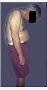
Case Study # 2:
ID/CC A 23 year old man presents with bilateral conjunctivitis, painful swelling of the right knee, bilateral heel pain, and painless ulcers on his penis.HPI He was diagnosed and treated for nongonococcal urethritis one week ago.PE Bilateral conjunctivitis with anterior uveitis (see pics); balanitis (shaft and glans penis are inflamed and scaly); arthritis of right knee and ankle.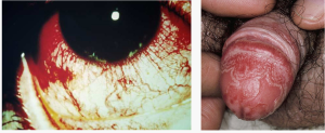 1. What is the Diagnosis?
Case Study # 3:
ID/CC A 40 year old white female complains of paleness and bluish discoloration of the hands, mainly upon exposure to cold, with redness upon rewarming, together with increasing pain in the knees, elbows, and hands over several months and recent difficulty swallowing solid food.HPI She also has mask-like facies with a limited range of expressionPE Smooth, shiny, tight skin over face and fingers; edema of hands and feet. (see pics).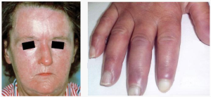
1. What is the Diagnosis?
Case Study # 4:
ID/CC A 70 year old male immigrant from England presents with pain in the right leg, producing an awkward gait, together with bilateral hearing loss.HPI He also has noted a progressive increase in hat sizePE Slight bowing of right tibia (see pic); normal rectal exam; mixed conductive and sensorineural hearing loss confirmed by audiometry.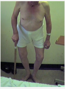 1. What is the Diagnosis?
Spot Diagnosis # 1:
A 2 year old with an autosomal dominant disorder.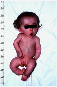 1. What is the Diagnosis?
Spot Diagnosis # 2:
Note the short limbs and the large vault of the skull.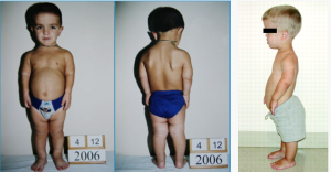
1. What is the Diagnosis?
Spot Diagnosis # 3:
Rare in the US, common in developing countries (& immigrants).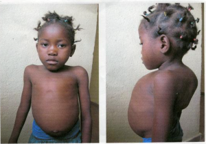 1. What is the Diagnosis?
-
Spot Diagnosis # 4:
Patient with a history of Rheumatoid Arthritis presented with painful left calf.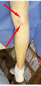 1. What is the Diagnosis?
Answers
CASE STUDY # 1:
ID/CC A 24 year old white male visits his family doctor complaining of low back pain and stiffness on active movement of the spine for almost a year; increasing in severity.
HPI The pain increases with movement and radiates down the posterior thigh, improving as the day progresses
PE Stooped posture; loss of lumbar lordosis and fixed kyphosis; tenderness over sacroiliac joints; reduced chest expansion; prominent abdomen (see the pic, it’s for an older pt with the same condition)
What is the Diagnosis?
ANSWER
Ankylosing Spondylitis – typical posture……. to puchae all the answers to the case studies click purchase below